Sarcomere
Smallest functional unit of striated muscle tissue.

A sarcomere (Greek σάρξ sarx "flesh", μέρος meros "part") is the smallest functional unit of striated muscle tissue. It is the repeating unit between two Z-lines. Sarcomeres give skeletal and cardiac muscle their striated appearance, which was first described by Van Leeuwenhoek. They are composed of long, fibrous proteins as filaments that slide past each other when a muscle contracts or relaxes.
- field
- Cell biology, muscle physiology
- known_for
- Smallest functional unit of striated muscle; sliding filament model of contraction
- structure
- Repeating unit between Z-lines; composed of actin (thin) and myosin (thick) filaments
- bands
- I-band, A-band, H-zone, M-line, Z-line
Lore & Background
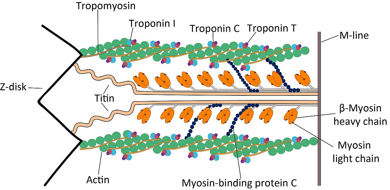
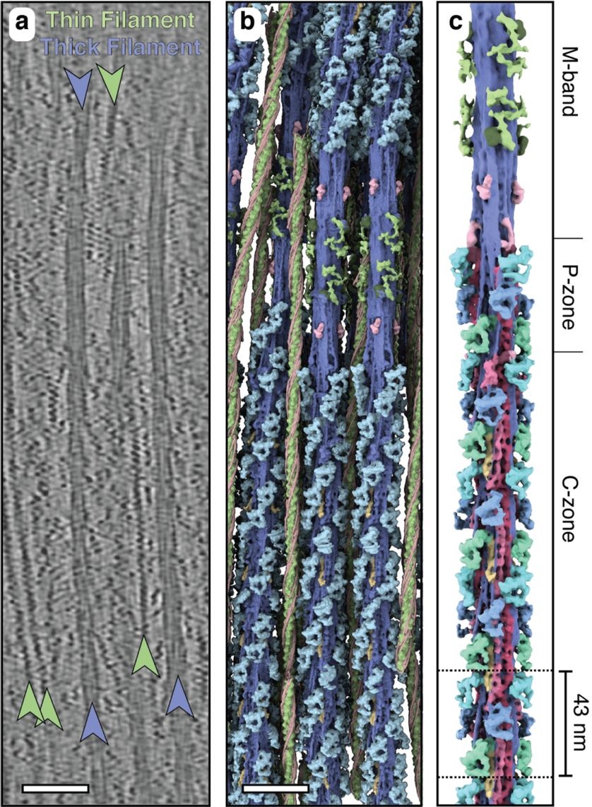
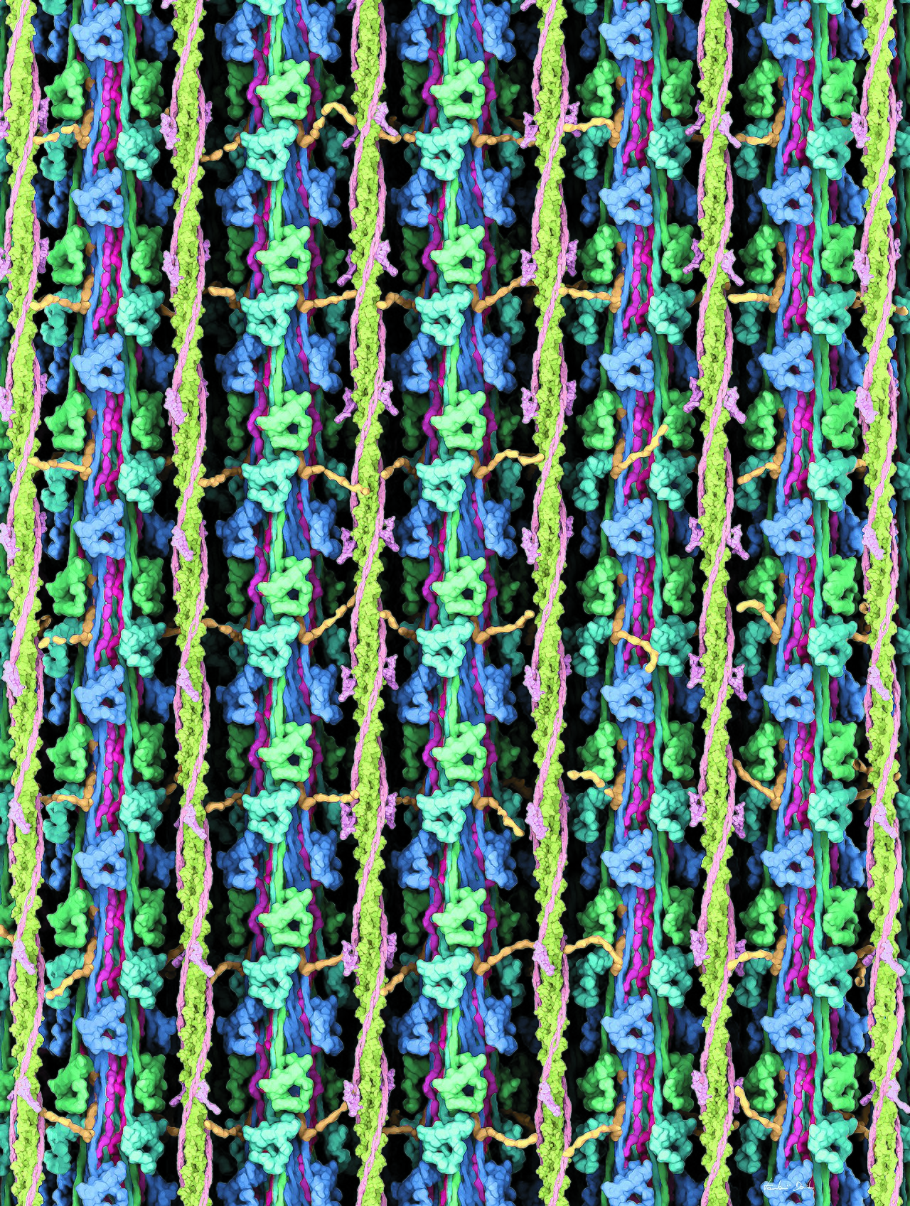
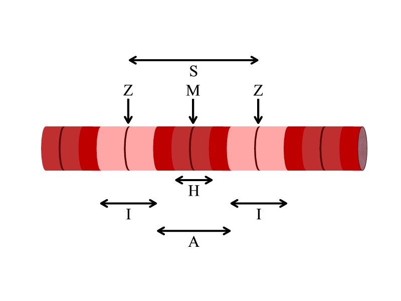
The sarcomere is defined as the segment between two neighbouring Z-lines (or Z-discs). In electron micrographs, the Z-line appears between the I-bands as a dark line that anchors the actin myofilaments. Surrounding the Z-line is the I-band (isotropic), the zone of thin filaments not superimposed by thick filaments. Following the I-band is the A-band (anisotropic), which contains the entire length of a single thick filament. Within the A-band is a paler region called the H-zone (from German "heller", brighter), the zone of thick filaments that has no actin. Within the H-zone is a thin M-line (from German "mittel", meaning middle), formed of cross-connecting elements of the cytoskeleton. Actin filaments are the major component of the I-band and extend into the A-band. Myosin filaments are bipolar and extend throughout the A-band, cross-linked at the centre by the M-band. The giant protein titin extends from the Z-line to the M-band, acting as a sarcomeric ruler. Another giant protein, nebulin, extends along the thin filaments and the entire I-band. The Z-line contains alpha-actinin, which cross-links actin filaments and titin molecules. The M-band contains myomesin and C-protein, which crosslink the thick filament system and the M-band part of titin. Muscle contraction occurs via the sliding filament model. Calcium ions bind with troponin C, altering tropomyosin structure to reveal cross-bridge binding sites on actin. The myosin head, bound to ATP, hydrolyzes it to ADP and inorganic phosphate, changing to a high-energy configuration. Upon binding to actin, the myosin head releases ADP and phosphate, returning to a low-energy configuration. A new ATP binds myosin, releasing actin. The A-bands do not change length during contraction, while I-bands and H-zone shorten.
Reader's Guide
The sarcomere is fundamental to understanding muscle contraction and the mechanical properties of striated muscle. Its repeating structure of actin and myosin filaments, organized into distinct bands (I-band, A-band, H-zone, M-line, Z-line), directly produces the sliding filament mechanism of contraction. The relationship between sarcomere structure and function is illustrated by the length-tension curve: force output decreases if the muscle is stretched so that fewer cross-bridges can form or compressed until actin filaments interfere. Vertebrates display a very limited range of sarcomere lengths, with roughly the same optimal length in all muscles of an individual and between species, whereas arthropods show tremendous variation (over seven-fold). The reasons for the lack of substantial sarcomere variability in vertebrates are not fully known. The sarcomere's components—including titin, nebulin, alpha-actinin, myomesin, and C-protein—provide a blueprint for assembly and stability. The interaction between actin and myosin in the A-band is responsible for muscle contraction, driven by ATP hydrolysis and regulated by calcium ions released from the sarcoplasmic reticulum via calcium-induced calcium release. Understanding the sarcomere is essential for fields ranging from exercise physiology to muscle disease research.
Did You Know?
- The sarcomere is the repeating unit between two Z-lines.
- Myosin can only bind to actin when the binding sites on actin are exposed by calcium ions.
- The giant protein titin extends from the Z-line to the M-band and is the biggest single highly elasticated protein found in nature.
- Upon muscle contraction, the A-bands do not change their length (1.85 micrometer in mammalian skeletal muscle), whereas the I-bands and the H-zone shorten.
The Architecture of the Repeating Unit
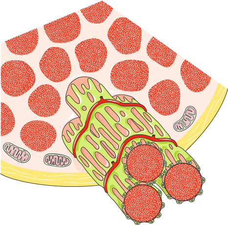
The sarcomere stands as the fundamental building block of striated muscle, defined as the segment stretching between two adjacent Z-discs. Within skeletal muscle, these units are nested inside tubular myofibrils, which themselves fill elongated muscle fibers generated during embryonic myogenesis. Under a microscope, the repeating sarcomeres produce the characteristic alternating pattern of dark and light bands that give striated muscle its name. At the core of each sarcomere lie two classes of fibrous protein filaments: the thick filaments built from myosin and the thin filaments composed of actin. Myosin presents a long fibrous tail capped by a globular head capable of gripping actin, and that same head also latches onto ATP to fuel movement. Actin filaments anchor directly to the Z-line, establishing the boundary of each unit. Critically, myosin can only engage actin once calcium ions have exposed the relevant binding sites. A separate structure called the costamere tethers the sarcomere to the outer membrane, the sarcolemma, integrating the contractile machinery with the cell's exterior.
The Band System and Its Nomenclature
The striated appearance of skeletal and cardiac muscle was first observed by Van Leeuwenhoek, and the pattern he saw is produced entirely by the orderly arrangement of sarcomeric bands. The Z-line, a term drawn from the German word zwischen meaning between, appears as a dark line in electron micrographs and serves as the anchor point for actin myofilaments. Flanking each Z-line lies the I-band, named for its isotropic optical behavior, which contains only thin filaments without any overlap from thick filaments. Beyond the I-band stretches the A-band, so called because of its anisotropic response under polarized light; this region spans the full length of a single myosin filament and houses both thick and thin filaments in parallel. Nested within the A-band is the paler H-zone, from the German heller meaning brighter, where thick filaments exist without any actin. At the very center of the sarcomere sits the M-line, derived from mittel meaning middle, formed by cross-connecting cytoskeletal elements. These German-origin names reflect the optical properties observed under polarization microscopy, and together they map the precise geometry of the contractile apparatus.
The Cascade of Contraction
Muscle contraction begins when a motor neuron releases acetylcholine into the neuromuscular junction, where it binds nicotinic receptors on the muscle cell surface. This triggers a sodium influx that fires an action potential, which propagates along transverse T-tubules until it reaches the sarcoplasmic reticulum. There, voltage-gated L-type calcium channels in the plasma membrane are activated and, in close physical association with ryanodine receptors on the sarcoplasmic reticulum, trigger a massive release of stored calcium ions—a process known as calcium-induced calcium release. The exact trigger, whether the physical opening of the L-type channel or the calcium itself, remains unresolved. Once free calcium floods the cytoplasm, it binds troponin C molecules scattered along tropomyosin, forcing tropomyosin to shift and expose the myosin-binding sites on actin. Myosin heads, previously locked in a low-energy state with ATP attached, can now form cross-bridges. During contraction the A-band holds steady at roughly 1.85 micrometers in mammalian skeletal muscle while the I-band and H-zone visibly shorten, drawing the Z-lines closer together. Relaxation occurs when calcium is actively pumped back into the sarcoplasmic reticulum.
Molecular Rulers and Structural Scaffolding
Beyond the headline actin and myosin, the sarcomere depends on an elaborate network of giant structural proteins that provide both elasticity and spatial organization. Titin, also called connectin, is the largest highly elasticated protein known in nature. It stretches from the Z-line, where it binds the myosin-based thick filament system, all the way to the M-band, where it is thought to interact with those same thick filaments. Titin serves as a molecular ruler and a blueprint guiding the assembly of the entire sarcomere, while simultaneously offering binding sites for numerous other proteins. A second giant, nebulin, is hypothesized to run along the thin filaments and the full length of the I-band, acting as a ruler for thin filament assembly. At the Z-disc, the protein alpha-actinin cross-links actin filaments to titin, while at the M-band, myomesin and C-protein tether the myosin system to titin's elastic portion. The M-line also hosts creatine kinase, an enzyme that regenerates ATP from ADP and phosphocreatine. Notably, smooth muscle cells lack this sarcomeric organization entirely, their myofibrils arranged without the repeating band pattern.
Gallery
Frequently Asked Questions
What exactly is a sarcomere?
It is the smallest contractile building block found in striated muscle fibers. Think of it as one repeating segment stretched between two Z-lines, and it is what gives skeletal and heart muscle their characteristic banded look.
What proteins make up a sarcomere?
Two main filament types do the work: thin actin filaments and thick myosin filaments. They interdigitate in a specific pattern to create the I-band, A-band, H-zone, and M-line regions visible under a microscope.
How does a sarcomere actually generate force?
Contraction happens through the sliding filament mechanism, where myosin heads grab onto actin and pull the thin filaments inward. This shortens the sarcomere and, repeated across thousands of units, produces a visible muscle contraction.
Who first noticed the striped pattern in muscle tissue?
Antonie van Leeuwenhoek was the first to describe the striated appearance of muscle under his microscope. The repeating sarcomere structure is what creates those alternating light and dark bands he originally observed.
Why is the sarcomere called the 'functional unit' of muscle?
Because a single sarcomere can independently shorten and generate tension, it is the smallest piece of muscle capable of doing mechanical work. Everything above it—myofibrils, whole fibers, entire muscles—is essentially a scaling-up of that one contractile unit.
More in Cell & Molecular Biology 1-16
Spotted an error? Know more?
This is a living reference — every entry is fact-audited, and reader corrections feed straight into our audit queue. Suggest an edit · See this site's audit record